Stampa:Anaplastic astrocytoma.jpg
Daqs tad-dehra proviżorja: 800 × 552 pixels. Riżoluzzjonijiet oħra: 320 × 221 pixels | 640 × 442 pixels | 1,024 × 707 pixels | 1,200 × 828 pixels
Fajl oriġinali (1,200 × 828 pixel, dimensjoni: 200 KB, tip ta' MIME: image/jpeg)
| Dan huwa fajl mill-Wikimedia Commons. Il-deskrizzjoni fuq il-paġna ta' diskussjoni oriġinali tidher hawn taħt.
|
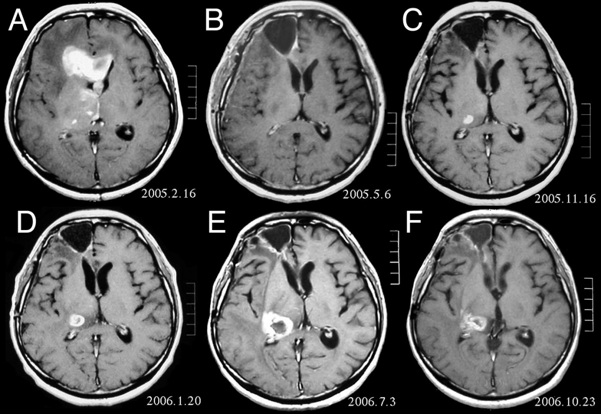
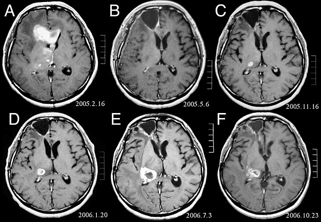
| Deskrizzjoni | MRI of brain. (A) Initial MRI on February 16, 2005, shows a tumor in the right and left frontal lobe as well as the right thalamus. (B) MRI after surgery, radiation and chemotherapy. The tumor has completely disappeared except for slight enhancement adjacent to the surgical margin. (C) Recurrence of the thalamic tumor despite maintenance chemotherapy on November 16, 2005. (D) Increase in size of the thalamic tumor two months after stereotactic radiotherapy. (E) After 6 cycles of TMZ therapy, the thalamic lesion enlarged, and the patient developed dysarthria and hemiparesis. (F) After 2 courses of treatment with interferon-beta and TMZ, the tumor shows a partial response. |
| Data | |
| Sors | Fujimaki T, Ishii H, Matsuno A, Arai H, Nakagomi T.Effectiveness of interferon-beta and temozolomide combination therapy against temozolomide-refractory recurrent anaplastic astrocytoma.World J Surg Oncol. 2007 Aug 4;5:89. PMID 17683572 doi:10.1186/1477-7819-5-89 |
| Awtur | Fujimaki T, Ishii H, Matsuno A, Arai H, Nakagomi T. |
| Permess (Użu mill-ġdid tal-fajl) |
BioMedCentral License |
Dan il-fajl huwa liċenzjat taħt it-termini tal-liċenzja Creative Commons Attribuzzjoni 2.0 Ġeneriku
- Inti ħieles:
- li taqsam – li tikkopja, tiddistribwixxi u tittrażmetti din l-opra
- li timmodifika – li tadatta l-biċċa xogħol
- Taħt il-kundizzjonijiet segwenti:
- attribuzzjoni – Għandek tattribwixxi x-xogħol bil-mod speċifikat mill-awtur jew minn min ta l-l-iċenzja (imma mhux b'xi mod li jissuġġerixxi ji jappoġjaw lilek jew l-użu tax-xogħol).
Kronoloġija tal-fajl
Agħfas fuq il-grupp data/ħin biex tara l-fajl biex tara kif jidher dak il-ħin.
| Data/Ħin | Minjatura | Qisien | Utent | Kumment | |
|---|---|---|---|---|---|
| kurrenti | 16:47, 25 Frar 2008 | | 1,200 × 828 (200 KB) | Filip em | {{Information |Description=MRI of brain. (A) Initial MRI on February 16, 2005, shows a tumor in the right and left frontal lobe as well as the right thalamus. (B) MRI after surgery, radiation and chemotherapy. The tumor has completely disappeared except f |
Użu tal-fajl
Il-Paġna segwenti twassal għal din l-istampa:
L-użu globali tal-fajl
Il-wikis segwenti jużaw dan il-fajl:
- Użu fuq ar.wikipedia.org
- Użu fuq bg.wikipedia.org
- Użu fuq cs.wikipedia.org
- Użu fuq da.wikipedia.org
- Użu fuq de.wikipedia.org
- Użu fuq el.wikipedia.org
- Użu fuq en.wikipedia.org
- Użu fuq es.wikipedia.org
- Użu fuq et.wikipedia.org
- Użu fuq hi.wikipedia.org
- Użu fuq hr.wikipedia.org
- Użu fuq hu.wikipedia.org
- Użu fuq it.wikipedia.org
- Użu fuq kk.wikipedia.org
- Użu fuq lb.wikipedia.org
- Użu fuq lv.wikipedia.org
- Użu fuq mk.wikipedia.org
- Użu fuq nl.wikipedia.org
- Użu fuq no.wikipedia.org
- Użu fuq pl.wikipedia.org
- Użu fuq ro.wikipedia.org
- Użu fuq sk.wikipedia.org
- Użu fuq sl.wikipedia.org
- Użu fuq sq.wikipedia.org
- Użu fuq sr.wikipedia.org
- Użu fuq uk.wikipedia.org